The LONVIA Edit
Curated insights on space, health, investment, and the future of living.
Circadian Science
Your eyes are not your clock
The LONVIA
Edit
Curated insights on space, health, investment, and the future of living.
Circadian Science
Your eyes are not your clock
8 min read
July 10, 2026
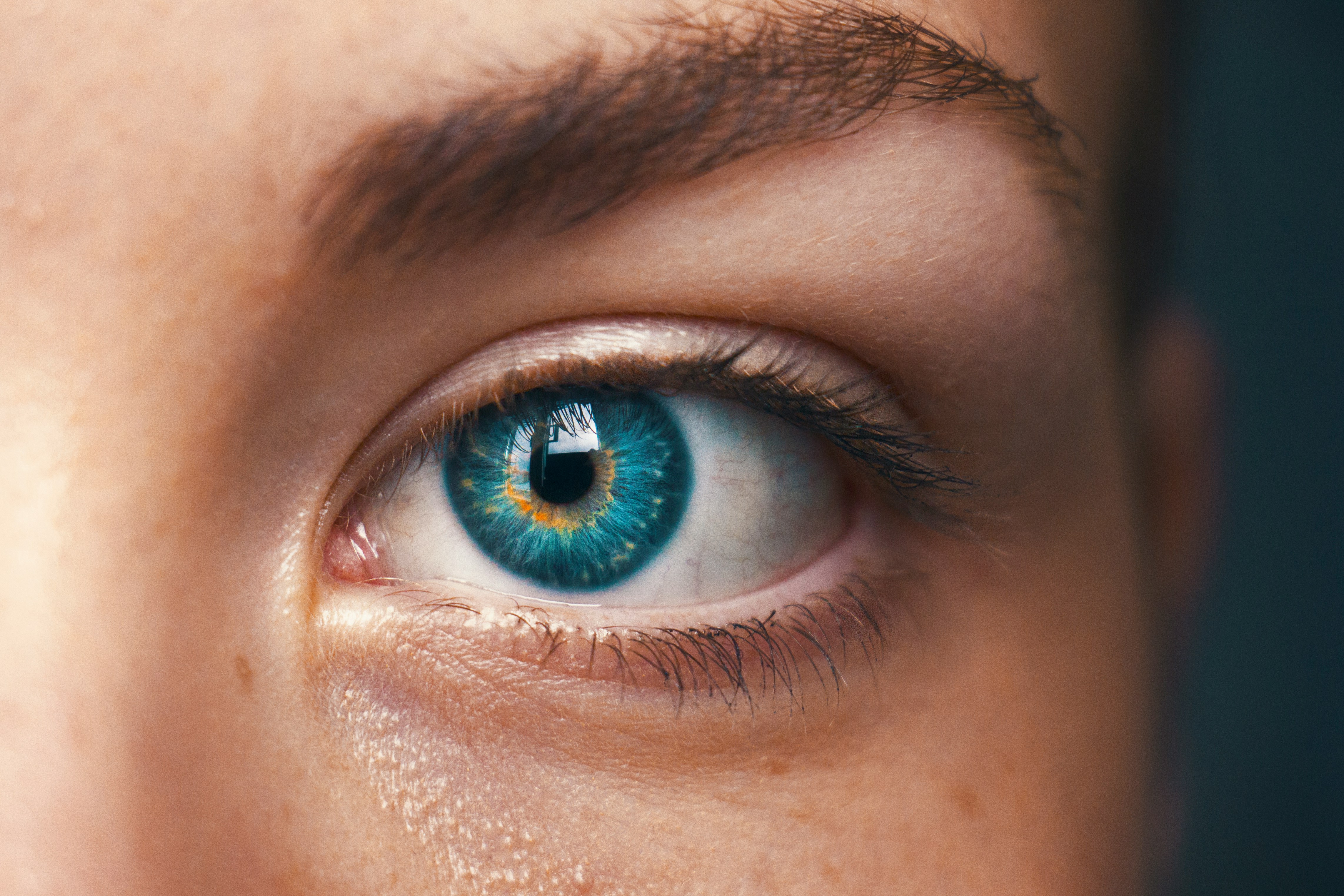
Every evening most of us perform the same small ritual. We turn the lights down, and the room softens. The eyes relax. The space reads as calm, as dim, as ready for sleep. In one sense this is exactly right. In another, it rests on an assumption that turns out to be false: that the part of the eye you see with and the part that tells your brain what time it is are the same thing, reading the same meter. They are not. They are two systems, sharing one window, measuring light in two different ways.
This is one of the more counter-intuitive results in modern human biology, and it is the foundation everything else about light and sleep is built on. It is worth setting out slowly.
What are the two jobs your eyes do?
Ask most people what the eye is for and they will say vision, which is correct as far as it goes. Light enters, lands on the retina, and is captured by rods and cones. Cones handle colour and daylight detail. Rods handle low light. Together they produce the sharp, moving, coloured picture you are using to read this sentence. The visual system is finely tuned, and its peak sensitivity sits in the green-yellow part of the spectrum, near 555 nanometres.
But the retina has a second job, quieter and much older in evolutionary terms. It reports how much light is in the environment, so the body can work out roughly where it is in the day. That job does not need a sharp image. It does not need colour or detail. It needs a slow, steady, reliable reading of ambient light, integrated over minutes and hours. And for most of the last century, nobody knew which cells were doing it.
Which cells in the eye set the circadian clock?
The textbook answer, for decades, was that rods and cones were the eye's only light detectors. Two lines of evidence eventually broke that picture apart.
The first came from blindness. In 1995 a group led by Charles Czeisler reported that some blind people, with no conscious perception of light at all, still showed the normal suppression of the sleep hormone melatonin when exposed to bright light. Their visual systems were gone. Their internal clocks were still receiving the message. Something in the eye was detecting light and passing it on, entirely outside of sight.
The second came from the laboratory. In 1999 a team led by Russell Foster showed that mice engineered to lack functional rods and cones could still reset their circadian rhythms to a light and dark cycle. Remove the entire visual apparatus, and the clock still keeps time by the sun.
The explanation arrived at the turn of the century. In 1998 Ignacio Provencio identified a new light-sensitive pigment and named it melanopsin. In 2002 David Berson and colleagues showed that a small population of retinal ganglion cells, the cells that normally just carry signals out of the eye, were themselves directly sensitive to light, because they contained it. These are the intrinsically photosensitive retinal ganglion cells, or ipRGCs. They make up only about 1 to 3 per cent of the retinal ganglion cells (Do and Yau, 2010). They are slow to respond, they form no image, and they measure one thing well: how much light is falling on the eye. That same year, Samer Hattar and colleagues traced their wiring and found it led to the suprachiasmatic nucleus, a cluster of neurons in the hypothalamus that acts as the body's master clock.
So a fraction of one per cent of what the retina does has nothing to do with vision, and it is wired directly to the structure that sets your circadian rhythm. You cannot feel it working. You have never noticed it. It has been running the whole time.
Why can two lights look the same but affect you differently?
Here is where it becomes practical. The visual system and this non-visual system do not respond to the same light in the same way. The visual system peaks near 555 nanometres, in the green-yellow. The melanopsin in the ipRGCs peaks lower, near 480 nanometres, in the short-wavelength blue region. Two different receptors, two different sensitivities.
The consequence is direct, and most people find it surprising. Two light sources can look equally bright to your eyes while delivering very different signals to your clock. Brightness, as you perceive it, is not a reliable guide to the biological weight of the light. The eye that sees and the eye that keeps time can disagree, and you have no conscious access to the disagreement.
How does light reach the body clock?
It helps to follow the message past the retina. When the ipRGCs detect light, they report it to the suprachiasmatic nucleus, which in turn governs the timing of melatonin release from the pineal gland. Melatonin is the body's night signal. It rises as the environment darkens and helps set the timing of sleep. Light reaching the ipRGCs at the wrong time holds that signal back. This is the mechanism behind the finding in blind patients: light suppresses melatonin because the pathway from eye to clock to pineal gland is intact, even when the pathway to sight is not. It is also why the effect is so easy to miss. Nothing about melatonin being suppressed produces a sensation. The room simply looks dim, and the biology quietly disagrees. In controlled work, Zeitzer and colleagues (2000) found the human clock responds to light at ordinary indoor levels, with roughly half the maximum melatonin effect reached near 100 lux.

What is melanopic EDI, and why was it needed?
If brightness is the wrong guide, the field needed a right one. In 2014 Robert Lucas and colleagues laid out the logic: light should be measured for each photoreceptor according to how that receptor actually responds, rather than forcing everything through the filter of human brightness perception. In 2018 this became an international standard, CIE S 026, which defined a set of melanopic quantities. The one that matters most here is melanopic equivalent daylight illuminance, usually shortened to melanopic EDI, or mEDI. It is light measured through the melanopsin filter rather than the visual one, expressed in the familiar units of lux so the numbers stay comparable. It answers the question brightness cannot: how much light is your circadian system actually receiving, at your eye.
Is daytime light as important as evening light?
With a metric in place, thresholds followed. In 2022 a large group of circadian scientists, led by Timothy Brown, published consensus recommendations for healthy adults: at least 250 melanopic lux at eye level during the day, no more than 10 in the evening, and no more than 1 while asleep. The evening ceiling gets most of the attention, but the daytime floor is there for a reason. The same system that should be quiet at night should be well fed during the day, and most indoor spaces deliver a fraction of the daytime figure, because rooms are lit for the visual system, which needs far less. Under-lit days and over-lit evenings are the same error in opposite directions: light specified for how it looks, not for what the second system receives.
What does this mean for your evening light?
The reason any of this matters is that it removes a judgement you thought you could make by looking. Dimming the lights in the evening is not wrong. It helps. But how dark a room feels is a report from your visual system, and your visual system is not the one keeping time. You can be sitting in what feels like a calm, dim, evening room while the receptor wired to your clock is still reading a signal much closer to daylight than you would guess. Nothing about the experience tells you. The only way to know is to measure the right quantity.
None of this is a claim that light is the only thing that governs sleep, or that hitting a number resolves everything. Sleep is shaped by many factors, people vary, and much of the underlying data comes from controlled laboratory conditions rather than living rooms. Honesty about those limits is part of taking the science seriously. But the central finding is not in doubt. There is a second light-sensing system in your eye, it does not care what the room looks like, and it has been setting your rhythm from a signal you cannot consciously feel. Once you know it is there, whether a room is dim enough stops being something you answer by looking, and becomes something you measure.
Frequently asked questions
Do your eyes control your body clock?
Not the visual part. A separate set of cells in the retina, containing the pigment melanopsin, detects overall light levels and signals the brain's master clock. They work independently of the rods and cones you see with.
What are ipRGCs?
Intrinsically photosensitive retinal ganglion cells. A small group of retinal cells, around 1 to 3 per cent of retinal ganglion cells, that respond directly to light and set circadian timing rather than forming images.
Can a room look dim but still affect my sleep?
Yes. How dark a room feels is judged by your visual system, which is not the one keeping time. The clock can register a significant light signal that you cannot consciously perceive.
What is melanopic EDI (mEDI)?
A measure of light weighted for the melanopsin system rather than for vision, expressed in lux and defined in the standard CIE S 026. It estimates what your circadian clock receives.
References
Czeisler CA, Shanahan TL, Klerman EB, et al. Suppression of melatonin secretion in some blind patients by exposure to bright light. New England Journal of Medicine. 1995;332(1):6-11.
Freedman MS, Lucas RJ, Soni B, et al. Regulation of mammalian circadian behavior by non-rod, non-cone, ocular photoreceptors. Science. 1999;284(5413):502-504.
Provencio I, Jiang G, De Grip WJ, et al. Melanopsin: an opsin in melanophores, brain, and eye. Proceedings of the National Academy of Sciences. 1998;95(1):340-345.
Berson DM, Dunn FA, Takao M. Phototransduction by retinal ganglion cells that set the circadian clock. Science. 2002;295(5557):1070-1073.
Hattar S, Liao HW, Takao M, Berson DM, Yau KW. Melanopsin-containing retinal ganglion cells: architecture, projections, and intrinsic photosensitivity. Science. 2002;295(5557):1065-1070.
Do MTH, Yau KW. Intrinsically photosensitive retinal ganglion cells. Physiological Reviews. 2010;90(4):1547-1581.
Gooley JJ, Rajaratnam SMW, Brainard GC, et al. Spectral responses of the human circadian system depend on the irradiance and duration of exposure to light. Science Translational Medicine. 2010;2(31):31ra33.
Zeitzer JM, Dijk DJ, Kronauer RE, Brown EN, Czeisler CA. Sensitivity of the human circadian pacemaker to nocturnal light: melatonin phase resetting and suppression. Journal of Physiology. 2000;526(3):695-702.
Lucas RJ, Peirson SN, Berson DM, et al. Measuring and using light in the melanopsin age. Trends in Neurosciences. 2014;37(1):1-9.
International Commission on Illumination. CIE S 026/E:2018. CIE System for Metrology of Optical Radiation for ipRGC-Influenced Responses to Light. Vienna: CIE; 2018.
Brown TM, Brainard GC, Cajochen C, et al. Recommendations for daytime, evening, and night-time indoor light exposure to best support physiology, sleep, and wakefulness in healthy adults. PLoS Biology. 2022;20(3):e3001571.


Ready to shape a healthier future?
Book an introductory conversation to explore how well-being can be integrated into your space or project
Ready to shape a healthier future?
Book an introductory conversation to explore how well-being can be integrated into your space or project
Ready to shape a healthier future?
Book an introductory conversation to explore how well-being can be integrated into your space or project
8 min read
July 10, 2026
Every evening most of us perform the same small ritual. We turn the lights down, and the room softens. The eyes relax. The space reads as calm, as dim, as ready for sleep. In one sense this is exactly right. In another, it rests on an assumption that turns out to be false: that the part of the eye you see with and the part that tells your brain what time it is are the same thing, reading the same meter. They are not. They are two systems, sharing one window, measuring light in two different ways.
This is one of the more counter-intuitive results in modern human biology, and it is the foundation everything else about light and sleep is built on. It is worth setting out slowly.
What are the two jobs your eyes do?
Ask most people what the eye is for and they will say vision, which is correct as far as it goes. Light enters, lands on the retina, and is captured by rods and cones. Cones handle colour and daylight detail. Rods handle low light. Together they produce the sharp, moving, coloured picture you are using to read this sentence. The visual system is finely tuned, and its peak sensitivity sits in the green-yellow part of the spectrum, near 555 nanometres.
But the retina has a second job, quieter and much older in evolutionary terms. It reports how much light is in the environment, so the body can work out roughly where it is in the day. That job does not need a sharp image. It does not need colour or detail. It needs a slow, steady, reliable reading of ambient light, integrated over minutes and hours. And for most of the last century, nobody knew which cells were doing it.
Which cells in the eye set the circadian clock?
The textbook answer, for decades, was that rods and cones were the eye's only light detectors. Two lines of evidence eventually broke that picture apart.
The first came from blindness. In 1995 a group led by Charles Czeisler reported that some blind people, with no conscious perception of light at all, still showed the normal suppression of the sleep hormone melatonin when exposed to bright light. Their visual systems were gone. Their internal clocks were still receiving the message. Something in the eye was detecting light and passing it on, entirely outside of sight.
The second came from the laboratory. In 1999 a team led by Russell Foster showed that mice engineered to lack functional rods and cones could still reset their circadian rhythms to a light and dark cycle. Remove the entire visual apparatus, and the clock still keeps time by the sun.
The explanation arrived at the turn of the century. In 1998 Ignacio Provencio identified a new light-sensitive pigment and named it melanopsin. In 2002 David Berson and colleagues showed that a small population of retinal ganglion cells, the cells that normally just carry signals out of the eye, were themselves directly sensitive to light, because they contained it. These are the intrinsically photosensitive retinal ganglion cells, or ipRGCs. They make up only about 1 to 3 per cent of the retinal ganglion cells (Do and Yau, 2010). They are slow to respond, they form no image, and they measure one thing well: how much light is falling on the eye. That same year, Samer Hattar and colleagues traced their wiring and found it led to the suprachiasmatic nucleus, a cluster of neurons in the hypothalamus that acts as the body's master clock.
So a fraction of one per cent of what the retina does has nothing to do with vision, and it is wired directly to the structure that sets your circadian rhythm. You cannot feel it working. You have never noticed it. It has been running the whole time.
Why can two lights look the same but affect you differently?
Here is where it becomes practical. The visual system and this non-visual system do not respond to the same light in the same way. The visual system peaks near 555 nanometres, in the green-yellow. The melanopsin in the ipRGCs peaks lower, near 480 nanometres, in the short-wavelength blue region. Two different receptors, two different sensitivities.
The consequence is direct, and most people find it surprising. Two light sources can look equally bright to your eyes while delivering very different signals to your clock. Brightness, as you perceive it, is not a reliable guide to the biological weight of the light. The eye that sees and the eye that keeps time can disagree, and you have no conscious access to the disagreement.
How does light reach the body clock?
It helps to follow the message past the retina. When the ipRGCs detect light, they report it to the suprachiasmatic nucleus, which in turn governs the timing of melatonin release from the pineal gland. Melatonin is the body's night signal. It rises as the environment darkens and helps set the timing of sleep. Light reaching the ipRGCs at the wrong time holds that signal back. This is the mechanism behind the finding in blind patients: light suppresses melatonin because the pathway from eye to clock to pineal gland is intact, even when the pathway to sight is not. It is also why the effect is so easy to miss. Nothing about melatonin being suppressed produces a sensation. The room simply looks dim, and the biology quietly disagrees. In controlled work, Zeitzer and colleagues (2000) found the human clock responds to light at ordinary indoor levels, with roughly half the maximum melatonin effect reached near 100 lux.
What is melanopic EDI, and why was it needed?
If brightness is the wrong guide, the field needed a right one. In 2014 Robert Lucas and colleagues laid out the logic: light should be measured for each photoreceptor according to how that receptor actually responds, rather than forcing everything through the filter of human brightness perception. In 2018 this became an international standard, CIE S 026, which defined a set of melanopic quantities. The one that matters most here is melanopic equivalent daylight illuminance, usually shortened to melanopic EDI, or mEDI. It is light measured through the melanopsin filter rather than the visual one, expressed in the familiar units of lux so the numbers stay comparable. It answers the question brightness cannot: how much light is your circadian system actually receiving, at your eye.
Is daytime light as important as evening light?
With a metric in place, thresholds followed. In 2022 a large group of circadian scientists, led by Timothy Brown, published consensus recommendations for healthy adults: at least 250 melanopic lux at eye level during the day, no more than 10 in the evening, and no more than 1 while asleep. The evening ceiling gets most of the attention, but the daytime floor is there for a reason. The same system that should be quiet at night should be well fed during the day, and most indoor spaces deliver a fraction of the daytime figure, because rooms are lit for the visual system, which needs far less. Under-lit days and over-lit evenings are the same error in opposite directions: light specified for how it looks, not for what the second system receives.
What does this mean for your evening light?
The reason any of this matters is that it removes a judgement you thought you could make by looking. Dimming the lights in the evening is not wrong. It helps. But how dark a room feels is a report from your visual system, and your visual system is not the one keeping time. You can be sitting in what feels like a calm, dim, evening room while the receptor wired to your clock is still reading a signal much closer to daylight than you would guess. Nothing about the experience tells you. The only way to know is to measure the right quantity.
None of this is a claim that light is the only thing that governs sleep, or that hitting a number resolves everything. Sleep is shaped by many factors, people vary, and much of the underlying data comes from controlled laboratory conditions rather than living rooms. Honesty about those limits is part of taking the science seriously. But the central finding is not in doubt. There is a second light-sensing system in your eye, it does not care what the room looks like, and it has been setting your rhythm from a signal you cannot consciously feel. Once you know it is there, whether a room is dim enough stops being something you answer by looking, and becomes something you measure.
Frequently asked questions
Do your eyes control your body clock?
Not the visual part. A separate set of cells in the retina, containing the pigment melanopsin, detects overall light levels and signals the brain's master clock. They work independently of the rods and cones you see with.
What are ipRGCs?
Intrinsically photosensitive retinal ganglion cells. A small group of retinal cells, around 1 to 3 per cent of retinal ganglion cells, that respond directly to light and set circadian timing rather than forming images.
Can a room look dim but still affect my sleep?
Yes. How dark a room feels is judged by your visual system, which is not the one keeping time. The clock can register a significant light signal that you cannot consciously perceive.
What is melanopic EDI (mEDI)?
A measure of light weighted for the melanopsin system rather than for vision, expressed in lux and defined in the standard CIE S 026. It estimates what your circadian clock receives.
References
Czeisler CA, Shanahan TL, Klerman EB, et al. Suppression of melatonin secretion in some blind patients by exposure to bright light. New England Journal of Medicine. 1995;332(1):6-11.
Freedman MS, Lucas RJ, Soni B, et al. Regulation of mammalian circadian behavior by non-rod, non-cone, ocular photoreceptors. Science. 1999;284(5413):502-504.
Provencio I, Jiang G, De Grip WJ, et al. Melanopsin: an opsin in melanophores, brain, and eye. Proceedings of the National Academy of Sciences. 1998;95(1):340-345.
Berson DM, Dunn FA, Takao M. Phototransduction by retinal ganglion cells that set the circadian clock. Science. 2002;295(5557):1070-1073.
Hattar S, Liao HW, Takao M, Berson DM, Yau KW. Melanopsin-containing retinal ganglion cells: architecture, projections, and intrinsic photosensitivity. Science. 2002;295(5557):1065-1070.
Do MTH, Yau KW. Intrinsically photosensitive retinal ganglion cells. Physiological Reviews. 2010;90(4):1547-1581.
Gooley JJ, Rajaratnam SMW, Brainard GC, et al. Spectral responses of the human circadian system depend on the irradiance and duration of exposure to light. Science Translational Medicine. 2010;2(31):31ra33.
Zeitzer JM, Dijk DJ, Kronauer RE, Brown EN, Czeisler CA. Sensitivity of the human circadian pacemaker to nocturnal light: melatonin phase resetting and suppression. Journal of Physiology. 2000;526(3):695-702.
Lucas RJ, Peirson SN, Berson DM, et al. Measuring and using light in the melanopsin age. Trends in Neurosciences. 2014;37(1):1-9.
International Commission on Illumination. CIE S 026/E:2018. CIE System for Metrology of Optical Radiation for ipRGC-Influenced Responses to Light. Vienna: CIE; 2018.
Brown TM, Brainard GC, Cajochen C, et al. Recommendations for daytime, evening, and night-time indoor light exposure to best support physiology, sleep, and wakefulness in healthy adults. PLoS Biology. 2022;20(3):e3001571.
Circadian Science
Your eyes are not your clock
8 min read
July 10, 2026
Every evening most of us perform the same small ritual. We turn the lights down, and the room softens. The eyes relax. The space reads as calm, as dim, as ready for sleep. In one sense this is exactly right. In another, it rests on an assumption that turns out to be false: that the part of the eye you see with and the part that tells your brain what time it is are the same thing, reading the same meter. They are not. They are two systems, sharing one window, measuring light in two different ways.
This is one of the more counter-intuitive results in modern human biology, and it is the foundation everything else about light and sleep is built on. It is worth setting out slowly.
What are the two jobs your eyes do?
Ask most people what the eye is for and they will say vision, which is correct as far as it goes. Light enters, lands on the retina, and is captured by rods and cones. Cones handle colour and daylight detail. Rods handle low light. Together they produce the sharp, moving, coloured picture you are using to read this sentence. The visual system is finely tuned, and its peak sensitivity sits in the green-yellow part of the spectrum, near 555 nanometres.
But the retina has a second job, quieter and much older in evolutionary terms. It reports how much light is in the environment, so the body can work out roughly where it is in the day. That job does not need a sharp image. It does not need colour or detail. It needs a slow, steady, reliable reading of ambient light, integrated over minutes and hours. And for most of the last century, nobody knew which cells were doing it.
Which cells in the eye set the circadian clock?
The textbook answer, for decades, was that rods and cones were the eye's only light detectors. Two lines of evidence eventually broke that picture apart.
The first came from blindness. In 1995 a group led by Charles Czeisler reported that some blind people, with no conscious perception of light at all, still showed the normal suppression of the sleep hormone melatonin when exposed to bright light. Their visual systems were gone. Their internal clocks were still receiving the message. Something in the eye was detecting light and passing it on, entirely outside of sight.
The second came from the laboratory. In 1999 a team led by Russell Foster showed that mice engineered to lack functional rods and cones could still reset their circadian rhythms to a light and dark cycle. Remove the entire visual apparatus, and the clock still keeps time by the sun.
The explanation arrived at the turn of the century. In 1998 Ignacio Provencio identified a new light-sensitive pigment and named it melanopsin. In 2002 David Berson and colleagues showed that a small population of retinal ganglion cells, the cells that normally just carry signals out of the eye, were themselves directly sensitive to light, because they contained it. These are the intrinsically photosensitive retinal ganglion cells, or ipRGCs. They make up only about 1 to 3 per cent of the retinal ganglion cells (Do and Yau, 2010). They are slow to respond, they form no image, and they measure one thing well: how much light is falling on the eye. That same year, Samer Hattar and colleagues traced their wiring and found it led to the suprachiasmatic nucleus, a cluster of neurons in the hypothalamus that acts as the body's master clock.
So a fraction of one per cent of what the retina does has nothing to do with vision, and it is wired directly to the structure that sets your circadian rhythm. You cannot feel it working. You have never noticed it. It has been running the whole time.
Why can two lights look the same but affect you differently?
Here is where it becomes practical. The visual system and this non-visual system do not respond to the same light in the same way. The visual system peaks near 555 nanometres, in the green-yellow. The melanopsin in the ipRGCs peaks lower, near 480 nanometres, in the short-wavelength blue region. Two different receptors, two different sensitivities.
The consequence is direct, and most people find it surprising. Two light sources can look equally bright to your eyes while delivering very different signals to your clock. Brightness, as you perceive it, is not a reliable guide to the biological weight of the light. The eye that sees and the eye that keeps time can disagree, and you have no conscious access to the disagreement.
How does light reach the body clock?
It helps to follow the message past the retina. When the ipRGCs detect light, they report it to the suprachiasmatic nucleus, which in turn governs the timing of melatonin release from the pineal gland. Melatonin is the body's night signal. It rises as the environment darkens and helps set the timing of sleep. Light reaching the ipRGCs at the wrong time holds that signal back. This is the mechanism behind the finding in blind patients: light suppresses melatonin because the pathway from eye to clock to pineal gland is intact, even when the pathway to sight is not. It is also why the effect is so easy to miss. Nothing about melatonin being suppressed produces a sensation. The room simply looks dim, and the biology quietly disagrees. In controlled work, Zeitzer and colleagues (2000) found the human clock responds to light at ordinary indoor levels, with roughly half the maximum melatonin effect reached near 100 lux.
What is melanopic EDI, and why was it needed?
If brightness is the wrong guide, the field needed a right one. In 2014 Robert Lucas and colleagues laid out the logic: light should be measured for each photoreceptor according to how that receptor actually responds, rather than forcing everything through the filter of human brightness perception. In 2018 this became an international standard, CIE S 026, which defined a set of melanopic quantities. The one that matters most here is melanopic equivalent daylight illuminance, usually shortened to melanopic EDI, or mEDI. It is light measured through the melanopsin filter rather than the visual one, expressed in the familiar units of lux so the numbers stay comparable. It answers the question brightness cannot: how much light is your circadian system actually receiving, at your eye.
Is daytime light as important as evening light?
With a metric in place, thresholds followed. In 2022 a large group of circadian scientists, led by Timothy Brown, published consensus recommendations for healthy adults: at least 250 melanopic lux at eye level during the day, no more than 10 in the evening, and no more than 1 while asleep. The evening ceiling gets most of the attention, but the daytime floor is there for a reason. The same system that should be quiet at night should be well fed during the day, and most indoor spaces deliver a fraction of the daytime figure, because rooms are lit for the visual system, which needs far less. Under-lit days and over-lit evenings are the same error in opposite directions: light specified for how it looks, not for what the second system receives.
What does this mean for your evening light?
The reason any of this matters is that it removes a judgement you thought you could make by looking. Dimming the lights in the evening is not wrong. It helps. But how dark a room feels is a report from your visual system, and your visual system is not the one keeping time. You can be sitting in what feels like a calm, dim, evening room while the receptor wired to your clock is still reading a signal much closer to daylight than you would guess. Nothing about the experience tells you. The only way to know is to measure the right quantity.
None of this is a claim that light is the only thing that governs sleep, or that hitting a number resolves everything. Sleep is shaped by many factors, people vary, and much of the underlying data comes from controlled laboratory conditions rather than living rooms. Honesty about those limits is part of taking the science seriously. But the central finding is not in doubt. There is a second light-sensing system in your eye, it does not care what the room looks like, and it has been setting your rhythm from a signal you cannot consciously feel. Once you know it is there, whether a room is dim enough stops being something you answer by looking, and becomes something you measure.
Frequently asked questions
Do your eyes control your body clock?
Not the visual part. A separate set of cells in the retina, containing the pigment melanopsin, detects overall light levels and signals the brain's master clock. They work independently of the rods and cones you see with.
What are ipRGCs?
Intrinsically photosensitive retinal ganglion cells. A small group of retinal cells, around 1 to 3 per cent of retinal ganglion cells, that respond directly to light and set circadian timing rather than forming images.
Can a room look dim but still affect my sleep?
Yes. How dark a room feels is judged by your visual system, which is not the one keeping time. The clock can register a significant light signal that you cannot consciously perceive.
What is melanopic EDI (mEDI)?
A measure of light weighted for the melanopsin system rather than for vision, expressed in lux and defined in the standard CIE S 026. It estimates what your circadian clock receives.
References
Czeisler CA, Shanahan TL, Klerman EB, et al. Suppression of melatonin secretion in some blind patients by exposure to bright light. New England Journal of Medicine. 1995;332(1):6-11.
Freedman MS, Lucas RJ, Soni B, et al. Regulation of mammalian circadian behavior by non-rod, non-cone, ocular photoreceptors. Science. 1999;284(5413):502-504.
Provencio I, Jiang G, De Grip WJ, et al. Melanopsin: an opsin in melanophores, brain, and eye. Proceedings of the National Academy of Sciences. 1998;95(1):340-345.
Berson DM, Dunn FA, Takao M. Phototransduction by retinal ganglion cells that set the circadian clock. Science. 2002;295(5557):1070-1073.
Hattar S, Liao HW, Takao M, Berson DM, Yau KW. Melanopsin-containing retinal ganglion cells: architecture, projections, and intrinsic photosensitivity. Science. 2002;295(5557):1065-1070.
Do MTH, Yau KW. Intrinsically photosensitive retinal ganglion cells. Physiological Reviews. 2010;90(4):1547-1581.
Gooley JJ, Rajaratnam SMW, Brainard GC, et al. Spectral responses of the human circadian system depend on the irradiance and duration of exposure to light. Science Translational Medicine. 2010;2(31):31ra33.
Zeitzer JM, Dijk DJ, Kronauer RE, Brown EN, Czeisler CA. Sensitivity of the human circadian pacemaker to nocturnal light: melatonin phase resetting and suppression. Journal of Physiology. 2000;526(3):695-702.
Lucas RJ, Peirson SN, Berson DM, et al. Measuring and using light in the melanopsin age. Trends in Neurosciences. 2014;37(1):1-9.
International Commission on Illumination. CIE S 026/E:2018. CIE System for Metrology of Optical Radiation for ipRGC-Influenced Responses to Light. Vienna: CIE; 2018.
Brown TM, Brainard GC, Cajochen C, et al. Recommendations for daytime, evening, and night-time indoor light exposure to best support physiology, sleep, and wakefulness in healthy adults. PLoS Biology. 2022;20(3):e3001571.